Ultrasound Findings: Gynaecological Edition!
One of our most popular newsletters was the one I wrote on ultrasound findings on liver and gallbladder ultrasound. As half of the population is women, gynae ultrasounds unsurprisingly constitute the vast majority of outpatient scanning.
One of our most popular newsletters was the one I wrote on ultrasound findings on liver and gallbladder ultrasound. As half of the population is women, gynae ultrasounds unsurprisingly constitute the vast majority of outpatient scanning.
It is the first port of call for patients presenting with complex menorrhagia, pelvic pain and emergency presentations. Hopefully this newsletter can you help you appreciate the types of findings on ultrasound scans after your referral. So here we go!
Case 1
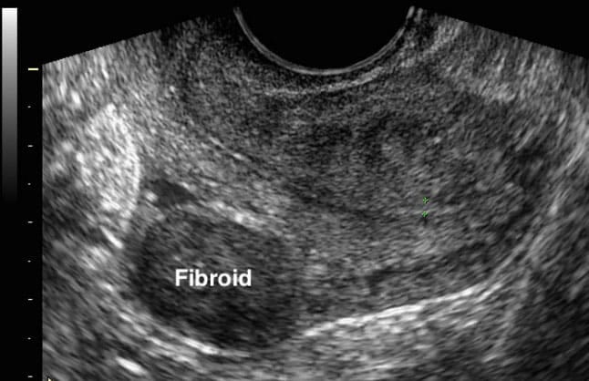
Mrs. A is a 42-year-old woman presenting with heavy menstrual bleeding for the past 8 months. Her periods are regular but prolonged, lasting up to 10 days, with passage of clots. She reports increasing pelvic pressure and a sensation of abdominal fullness but denies dysmenorrhea or intermenstrual bleeding. There is no history of postcoital bleeding. Examination reveals a palpable, irregularly enlarged uterus corresponding to a 14-week size.You decide to order an ultrasound.
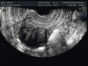
Ultrasound appearance: Well-defined, solid, often hypoechoic masses within the uterine wall. They can distort the uterine contour or cavity.
Uterine fibroids are benign smooth muscle tumors of the uterus that can vary in size and number. They may grow in different locations: within the uterine wall (intramural), beneath the lining (submucosal), or on the outer surface (subserosal). Fibroids can cause heavy menstrual bleeding, pelvic pressure, or may be asymptomatic. Treatment options broadly include:
- Medical management: NSAIDs, tranexamic acid, hormonal therapies (e.g., combined oral contraceptives, levonorgestrel-releasing intrauterine system) for symptom control.
- Minimally invasive procedures: Uterine artery embolisation, hysteroscopic myomectomy for submucosal fibroids.
- Surgical options: Myomectomy or hysterectomy for women with significant symptoms or when fertility is not a concern.
Case 2
Mrs. B is a 62-year-old postmenopausal woman presenting with painless vaginal bleeding for the past 3 weeks. She has no history of hormone replacement therapy and reports unintentional weight loss and fatigue. There is no history of pelvic pain or discharge. Examination reveals a normal-sized uterus with no palpable adnexal masses.
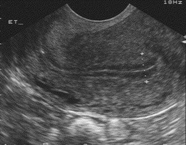
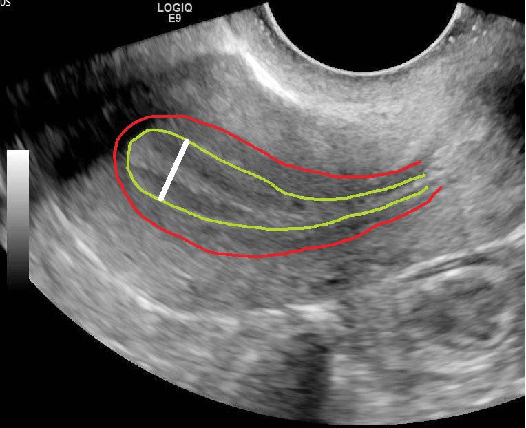
Ultrasound appearance: Endometrial thickness exceeding the expected range for cycle phase, or with irregular echotexture.
This is a thickened endometrium and needs to be investigated with hysteroscopy. There is a high risk that this patient may have an underlying cancer.
Case 3
Miss C is a 24-year-old woman presenting with intermittent lower abdominal pain for 2 months, sometimes associated with bloating. She reports regular menstrual cycles with no abnormal bleeding. There is no history of fever, gastrointestinal symptoms, or weight loss. On examination, the pelvis is soft with mild tenderness in the right adnexa, and no masses are palpable. You decide to perform an ultrasound scan:
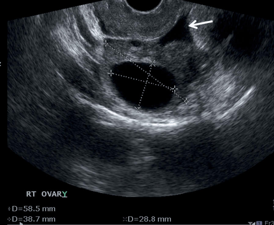
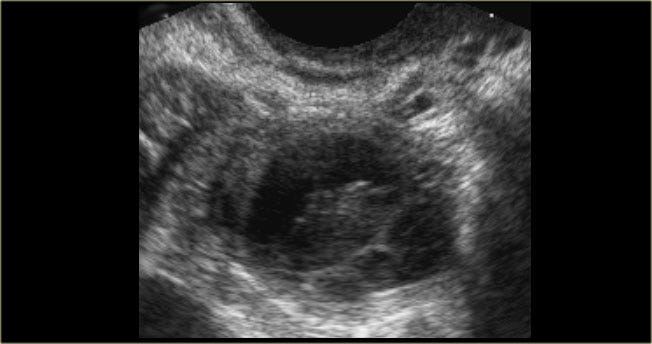
This first scan shows a simple ovarian cyst measuring almost 6cm. It is thin-walled, anechoic, no internal echoes. As it is a simple cyst, it will require a follow up scan to see if there is any change in its size or nature.
The second cyst shows a complex cyst which requires urgent referral.
Case 4:
Miss D is a 28-year-old woman presenting with sudden-onset lower abdominal pain and spotting per vagina for 1 day. Her last menstrual period was 6 weeks ago, and a home pregnancy test was positive. She reports no fever or gastrointestinal symptoms, but describes dizziness and mild shoulder tip pain. On examination, she is tender in the left lower quadrant with mild guarding. On performing an ultrasound you find:
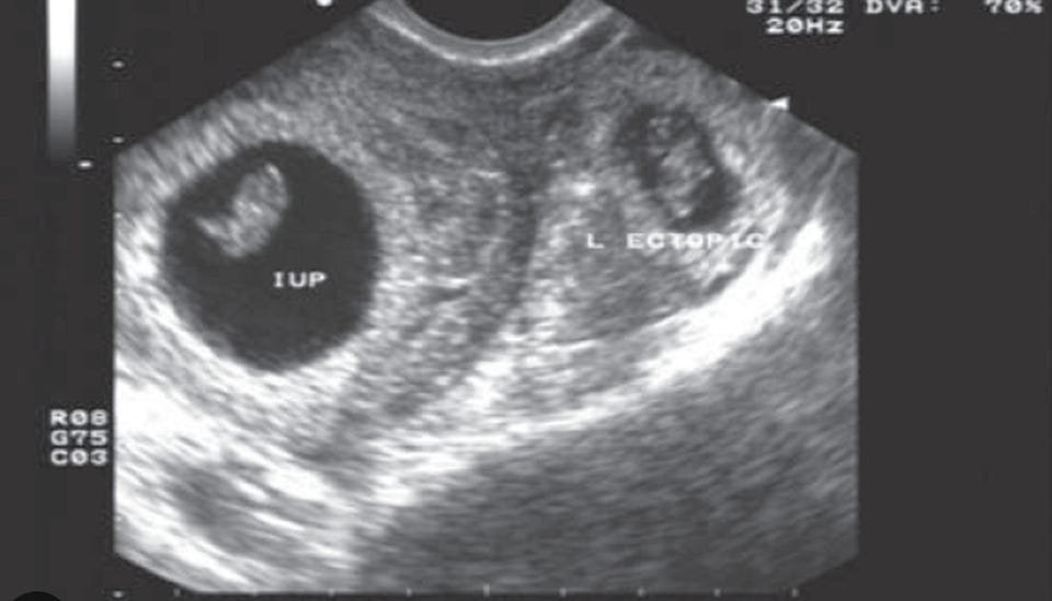
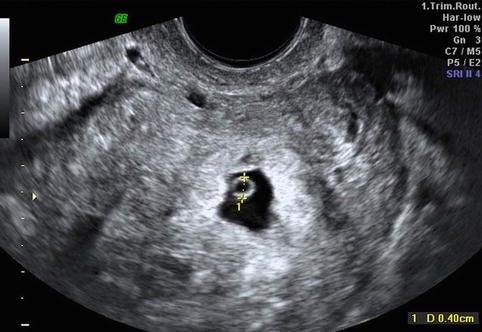
Ultrasound findings: intrauterine mass with an adnexal mass and free fluid in the pelvis.
This is a concerning scan as there is an intrauterine pregnancy as well as an ectopic pregnancy. In most scenarios, it depends on how unwell the patient is as well as the B-HCG test. Broadly speaking, the options include:
- Medical: Methotrexate for stable, unruptured ectopics.
- Surgical: Laparoscopic salpingectomy or salpingostomy for ruptured or high-risk cases.
- Expectant: Hemodynamic stabilisation and close follow-up of β-hCG levels
Case 5:
Miss E is a 30-year-old woman presenting with intermittent lower abdominal pain and irregular light vaginal bleeding for 3 weeks. Her menstrual cycles are usually regular, and she denies missed periods or nausea. She has no significant medical history and takes no hormonal contraception. On examination, the abdomen is soft with mild tenderness in the left adnexa, and there are no palpable masses. You perform an ultrasound which shows the following:
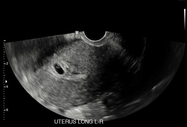
Ultrasound appearance: Gestational sac with yolk sac or fetal pole within the uterus
What a surprise! Trust me.. it happens. This patient needs to be told the news and subsequently referred to the midwife. Thankfully, the pregnancy is in the uterus and the symptoms are likely down to this.
So.. those were some scans from common gynaecology presentations.
Pelvic ultrasound provides important clarity in gynaecological care. transforming uncertainty into actionable diagnosis. Understanding what happens beyond the referral helps us manage patient expectations, streamline care, and communicate more effectively with radiology colleagues. It helps to set expectations and helps patients understand what will happen next.
I hope this has informed you about the role of gynaecological investigations.
Until next time!
Welcoming October… with some FRESH CPD
October is here and 2025 will be over in a blink.
Time isn't the only thing moving fast - it’s also knowledge which makes it all the important to keep up to date with your CPD.
We have TWO fantastic events planned for you all. Some of these include:
- ECG Masterclass - Sunday 12th October- Join us this SUNDAY for our popular ECG Masterclass. It is the perfect way to learn or revise ECG interpretation with plenty of explanations, reference images and examples to apply your knowledge. If you book onto this, you will also receive a FREE ECGs Made Easy e-book, authored by the Pareto Team.You can book here
Remember - if you can’t make the live event, you will also receive a recording to watch at a later date!
- Minor Injury Masterclass - Sunday 19th November 2025 - Where there is illness, there is also injury and this is exactly why we’ve organised this brilliant course. After the success of our minor injuries and urgent care courses, the last point in the triangle was minor injuries. Join us as we explore the world of injuries including MSK, soft tissue, burns and tricky areas such as nose and eyes. We will explore areas such as wound management and referral criteria if the injury isn’t “minor.” Book now!

Remember - if you can’t make the live event, you will also receive a recording to watch at a later date!
Website: www.paretoeducation.co.uk
Instagram: www.instagram.com/pareto_ed
Twitter: www.twitter.com/pareto_ed
Youtube: https://bit.ly/3DPm23c
Email: info@paretoeducation.co.uk
Originally published on Substack.